

Tag: blood test

  • Understanding Ovarian Cancer: Early Detection, Genetic Testing, and Prevention

    Understanding Ovarian Cancer: Early Detection, Genetic Testing, and Prevention

    Every day, 11 women in the UK lose their lives to ovarian cancer. This disease claims more lives than all other gynaecological cancers combined. Tragically, most cases are diagnosed at an advanced stage—stage 3 or 4—where the survival rate for stage 4 is a mere 15%. However, if detected early, at stage 1, the five-year survival rate soars to 95%.

    Ovarian cancer is shrouded in myths and misconceptions, leading to a lack of awareness among both women and healthcare professionals about effective prevention and early detection methods. With advancements in science, we now have the tools to detect this disease early and, in some cases, prevent it altogether, significantly improving survival rates.

    This video is essential viewing for anyone with a family history of ovarian cancer, anyone with ovaries, or anyone who cares about someone with ovaries. Join us as we dispel myths, spread awareness, and empower you with life-saving knowledge about ovarian cancer.

    Genetics of Ovarian Cancer

    Let’s first talk about the genetics of ovarian cancer. 

    About 20% of ovarian cancers are caused by a genetic variant inherited from a parent. You might have heard people describing this as having inherited a “gene mutation” or “faulty gene”. 

    The most common genes involved in this are BRCA1 Or B-R-C-A-1 and BRCA2. 

    If you have inherited a harmful copy of one of these genes, we medically call this having a “pathological” variant. For patients, this might mean your risk of ovarian cancer might be up to 60% in your lifetime. BRCA1 and 2 are not the only genes which are linked to ovarian cancer, there are other genes which are less common and carry different risks. 

    Angelina Jolie famously shared her story of finding out about her BRCA 1 mutation in 2013 in the New York Times.  I think she really helped to open the conversation around genetic screening for lots of women. She had sadly already lost her mother to cancer, who was diagnosed with breast and ovarian cancer. Angelina Jolie chose to have risk-reducing surgery.  Since then many other women have followed in her footsteps, talking about their genetic mutations and surgical decisions such as Christina Applegate, Sharon Osborne and many not so close to Hollywood like me.

    So who should have genetic screening?

    I want to share the words of Dr Mary-Claire King, she is the scientist who first identified the BRCA1 gene in 1990. 

    She says “To identify a woman as a carrier only after she develops cancer is a failure of cancer prevention,”.

    I whole heartedly agree with her. Despite the passing of decades since the discovery of the BRCA1 gene far too many people only find out they carry a gene after a cancer diagnosis in themselves or someone they love.

    Over 95% of women who have inherited a harmful variant in BRCA1 or BRCA2 are undiagnosed in the UK. That’s hundreds of thousands of people who don’t know they are at high risk of cancer, ovarian cancer, plus breast cancer and more. 

    I am extremely fortunate to have been able to access private genetic screening. And I am passionate about bringing this option to more patients and am proud we are able to offer this at Coyne Medical. We can take a sample in clinic after talking through the options, we usually test for multiple cancer genes linked to several cancers including ovarian, breast, colon and more. We only test for genetic changes which are linked to proactive steps you can take to reduce your risk. Such as recommendations for extra screening or checks. 

    But I know that is not an option for all patients, so I was also delighted that in March this year, the guidelines on NHS funded testing were updated by NICE. They now recommend that anyone who has a family history of ovarian cancer in a first or second degree relative be considered for genetic testing.

     A first-degree relative is for instance parent or sister. A second-degree relative is much broader and includes grandparents, aunts, nieces or half-sisters. This includes relatives on either your mum or dad’s side of the family.  So if private testing is not an option for you do speak to your GP about the new guidelines. In the past it may be that you were denied testing on the NHS but could now be eligible.

    What if you don’t have any family history of ovarian cancer?

    Yes, you can still have genetic screening. Traditionally we have only screened patients with a family history. We know from large studies on breast and ovarian cancer that if we only use the family history criteria we will miss over 50% of people with a pathological mutation in BRCA1 or BRCA2.  So if you really want toif you want to get serious about optimising your cancer screening and prevention we think genetic screening is crucial. You don’t want to be one of the patients that only find out after they get cancer, knowing you might have been able to prevent it earlier. 

    Personally, I know the truth behind the statistics. I tested positive for a pathological variant in a gene called PALB2 in 2021. It puts me at increased risk of ovarian cancer and breast cancer. I did not meet the traditional criteria for genetic testing and would not have known without choosing to have private genetic screening. I feel incredibly lucky I found my gene mutation while I was completely healthy, instead of after having a cancer. I have been able to join Angelina Jolie in choosing preventative risk-reducing surgery. To know my risk and then take action to reduce my risk of cancer feels like a very special gift. I would love to see more women share this. 

    Could a family history of other cancers be linked to ovarian cancer?

    Yes, many of the genes that increase the risk of ovarian cancer are linked to other cancers.

    If your family history includes breast cancer, pancreatic cancer or prostate cancer. Those are all flags that you have a higher chance of having inherited a gene mutation. We would certainly encourage you to consider genetic screening. 

    And it’s not just women. We would also encourage men with a family history of these cancers to consider genetic screening. Finding a gene mutation will help us manage their health risks and cancer screening. But it will also potentially help their daughters, granddaughters, nieces, sisters…

    MYTH is that: “It doesn’t matter as there is no family history on my Mum’s side.”

    Wrong. You can inherit these gene mutations from either your mother or father. 

    Women who inherit from their Dad are more often missed. Men are less likely to get breast cancer and can’t get ovarian cancer. So for their daughter their family history often appears less worrying. The same is true for people from small families. A history of cancer in a grandmother or aunt on Dad’s side of the family, is often not talked about or not seen as a big risk factor. I would encourage everyone to find out the most they can about their family history. During the dark days of the first lockdown I started researching my family tree which has been fascinating. After I discovered that I had a gene mutation I searched in more detail and got a copy of my great grandmother’s death certificate, this showed she had died from breast cancer in her 40’s. I almost certainly inherited my mutation from her. So get talking to relatives if you can or use the wonders of the internet to do a bit more research. Of course, we focussed on ovarian cancer today but family history of all cancers as well as other diseases such as heart attacks are also really important information. 

    Do you have any Jewish ancestry?

    Having a parent or grandparent of Jewish ancestry is a big risk factor for inheriting a BRCA1 or BRCA2 genetic variant, up to a 1 in 40 chance. We would recommend all people with this risk factor use genetic screening to find out if they carry a high-risk mutation. The NHS has recently started a project offering testing for gene variants in only the BRCA1 and BRCA2 genes, as these are the genes which are more commonly mutated in those with Jewish ancestry, if you’re interested you can find out more about the project online and I’ll put the link to that in our notes. 

    How does finding a high-risk ovarian cancer gene help you prevent cancer?

    Some women will opt to have their ovaries and fallopian tubes removed. Especially if they have already entered menopause, you could say they are no longer serving any purpose so why keep them and add to your cancer risk? 

    The decision can be more difficult for younger women, for whom surgery will bring on an early menopause and impact on their choices for fertility. It is important that women in this scenario get the best advice on which option is best for them. And their options for pregnancy in the future. 

    There is a brilliant UK study called “Protector”, it is led by  Professor Ranjit Manchanda, who is a great advocate for increasing access to genetic screening for women. The study offers women who want to keep their ovaries the option of removing their fallopian tubes only initially, followed by removal of the ovaries at a later date. We know many ovarian cancers start at the end of the fallopian tubes where they meet the ovaries. The study wants to show this option can help women who wish to delay their surgery, so this option could help reduce risk without the negative impact of menopause in younger women. Both surgical options are big decisions, and need careful thinking through the pros and cons so each woman can find the right decision for her health. Thankfully the actual surgeries are usually laparoscopic or keyhole operations, and can usually be done as a daycase procedure. 

    Can I do anything else to prevent ovarian cancer?

    Yes, there are lots of factors we can focus on to reduce the risk of ovarian cancer. And they will also reduce your risk of lots of other cancers such as breast and colon cancer. Keeping a healthy weight and not smoking are the two biggest risk factors which you can control. The combined oral contraceptive pill also reduces risk, even if only taken for a short period of time. The protection from taking the pill also persists, so the positive benefit continues for several years after stopping the pill. For high-risk women though such as having a BRCA1 or 2 mutation the combined oral contraceptive increases the risk of breast cancer so it may not be suitable as an option. Breastfeeding is protective for ovarian cancer risk, with a 24% risk reduction. I know personally breastfeeding can be really challenging, certainly the hardest part of life as a new mum with my first baby. So I am really conscious of not adding to the mummy guilt with this but if you have a family history it is good to know that this choice could have a positive impact on your own health.  Having children reduces the chance of having ovarian cancer. Of course not alone a reason to have a baby! There has been a lot of concern about using talcum powder on the genital area and ovarian cancer, the evidence is mixed but it is sensible to avoid this. 

    Early Detection

    But now let’s think about early detection. For those cancers we can’t prevent, we need to diagnose them as early as possible. Early diagnosis is vital. Sadly two-thirds of women are diagnosed at a late stage.

    If we can diagnose women early at stage 1 the survival rate is over 94% at 5 years, versus only 16% for women diagnosed at stage 4. So how can we screen you for ovarian cancer and detect it early?

    For women who do not have a high-risk gene mutation we don’t routinely recommend all women using ultrasound or the CA125 blood test for ovarian cancer screening because the biggest UK trial didn’t show this could improve survival in women. They studied over 200,000 women over about 16 years, split into 3 groups, no screening, screening with an ultrasound scan and screening with an ultrasound scan and a blood test for CA125. The results showed women screened with an ultrasound and the CA125 blood test did seem to be diagnosed with cancer at an earlier stage, but they couldn’t show this saved a significant number of lives. We know the CA125 blood test is not perfect, it can give “false alarms”, as other conditions like endometriosis or fibroids can cause high CA125 levels too.  

    But the results were different in women who have are at “high risk” such as with a BRCA gene mutation, they are now recommended to consider regular screening with CA125 blood test and a special algorithm for monitoring their results called ROCA. The test has been available privately for some time but this is a new recommendation in 2024 for the NHS and not yet easily available nationally but hopefully this will change soon. The ROCA test has been show to detect ovarian cancer earlier, before symptoms appear. For high-risk women they can have this done 3 times a year, the results will be tracked and if a significant increase is noted this will prompt more tests. This might be a good option for high-risk gene carriers to consider, especially if you have decided against surgery or are waiting to have done at an older age. 

    Are there are any other ways of screening for ovarian cancer?

    Yes we now have a new blood test that can be used to screen for ovarian cancer. It uses a normal blood sample taken in the clinic and the laboratory to look for ‘circulating tumour cells’, (CTCs). When a cancer or ‘tumour’ starts to grow in the body some of the cells will get into the bloodstream called CTCs. The really exciting part of this is that these CTCs can be found in early-stage 1 cancers.  

    The blood test is called a Multi Cancer Early Detection test, we offer tests by Trucheck that can either focus on female cancers, including breast and ovarian cancer,  or we can screen for up to 70 solid organ tumours. We offer this test at Coyne Medical, always after a full consultation with the doctor to understand if it’s the right test for you. We always want to make sure that patients are up to date with their standard cancer screening as these newer tests should be seen as a great optional add-on. 

    Likewise we also use whole-body MRI scans as an add-on cancer screening tool. These scans from from head through the neck, chest and abdomen to the pelvis. In the pelvic area they can detect cysts or fluid which can indicate an ovarian cancer. In large studies of whole-body MRI the rate of detecting a cancer is 1 to 2% in healthy adults over 40, but this has to be balanced against finding small ‘abnormalities’ which can cause stress and need further checks. That’s why it’s important you go through the possibilities with your doctor before testing. As well as making sure your doctor is there to support you through the results and any follow-up.

     MYTH: “A smear test checks for ovarian cancer.”

    This is sadly common, about 1 in 4 women think this is true but sadly not. Cervical screening will not detect ovarian cancer. Though it is a really important cancer screening to make sure you are up to date with. 

    Knowing the symptoms is key for every woman

    Symptoms to watch for include:

    • Persistent increase in abdominal size or bloating (not bloating that comes and goes)
    • Persistent pelvic and abdominal pain
    • Unexplained change in bowel habits
    • Difficulty eating and feeling full quickly, or feeling nauseous
    • Needing to urinate (wee) more regularly

    Other symptoms can include tiredness, losing weight, pain during sex, and back pain. These can be symptoms of many other conditions too so don’t panic but see your doctor. Your doctor might check a blood test called CA125 and you may also need an ultrasound scan. 

    Knowledge is Power

    Knowledge is power, yet many women and doctors are not well-informed about ovarian cancer. A study by Target Ovarian Cancer found that less than 5% of women felt confident recognizing its signs. Surprisingly, many doctors also mistakenly believe that symptoms only appear in the late stages of the disease. 

    We’re committed to empowering women with the knowledge they need to make informed health decisions. If you know other women who could benefit from this information, please share it with them. Together, we can spread awareness and equip more women with the knowledge they need.

    Follow us if you’d like to learn more about disease prevention and early detection. 

    Cancer Research UK [https://www.cancerresearchuk.org/health-professional/cancer-statistics/statistics-by-cancer-type/ovarian-cancer/mortality#heading-Zero] Accessed 8 May 2024.

     Chandrasekaran D, Manchandra R. ‘Germline and somatic genetic testing in ovarian cancer patients’. BJOG 2018: volume 125, issue 11, page 1,460. DOI: 10.1111/1471-0528.15225

    Evans, D.G., Shenton, A., Woodward, E. et al. Penetrance estimates for BRCA1 and BRCA2based on genetic testing in a Clinical Cancer Genetics service setting: Risks of breast/ovarian cancer quoted should reflect the cancer burden in the family. BMC Cancer 8, 155 (2008). https://doi.org/10.1186/1471-2407-8-155

    https://ascopost.com/issues/february-10-2015/dr-mary-claire-king-proposes-population-screening-in-all-young-women-for-brca-mutations/#:~:text=%E2%80%9CTo%20identify%20a%20woman%20as,Dr

    Manchanda R, Blyuss O, Gaba F, et al. Current detection rates and time-to-detection of all identifiable BRCA carriers in the Greater London population, Journal of Medical Genetics 2018;55:538-545.

    National Institute of Clinical Excellence. Ovarian cancer: identifying and managing familial and genetic risk, NICE Published 21/3/2024, [https://www.nice.org.uk/guidance/ng241] Accessed 8 May 2024.

    Beitsch PD, Whitworth PW, Hughes K, Patel R, Rosen B, Compagnoni G, Baron P, Simmons R, Smith LA, Grady I, Kinney M, Coomer C, Barbosa K, Holmes DR, Brown E, Gold L, Clark P, Riley L, Lyons S, Ruiz A, Kahn S, MacDonald H, Curcio L, Hardwick MK, Yang S, Esplin ED, Nussbaum RL. Underdiagnosis of Hereditary Breast Cancer: Are Genetic Testing Guidelines a Tool or an Obstacle? J Clin Oncol. 2019 Feb 20;37(6):453-460. doi: 10.1200/JCO.18.01631. Epub 2018 Dec 7. PMID: 30526229; PMCID: PMC6380523. https://www.ncbi.nlm.nih.gov/pmc/articles/PMC6380523/

    Petrucelli N, Daly MB, Pal T. BRCA1- and BRCA2-Associated Hereditary Breast and Ovarian Cancer. 1998 Sep 4 [Updated 2023 Sep 21]. In: Adam MP, Feldman J, Mirzaa GM, et al., editors. GeneReviews® [Internet]. Seattle (WA): University of Washington, Seattle; 1993-2024. Available from: https://www.ncbi.nlm.nih.gov/books/NBK1247/

    Ozanne EM, O’Connell A, Bouzan C, Bosinoff P, Rourke T, Dowd D, Drohan B, Millham F, Griffin P, Halpern EF, Semine A, Hughes KS. Bias in the reporting of family history: implications for clinical care. J Genet Couns. 2012 Aug;21(4):547-56. doi: 10.1007/s10897-011-9470-x. Epub 2012 Jan 12. PMID: 22237666.

    Metcalfe KA, Eisen A, Lerner-Ellis J, Narod SA. Is it time to offer BRCA1 and BRCA2 testing to all Jewish women? Curr Oncol. 2015 Aug;22(4):e233-6. doi: 10.3747/co.22.2527. PMID: 26300672; PMCID: PMC4530819.

    The NHS Jewish BRCA Testing Programme

    The Protector Study

    Cancer Research UK, [https://www.cancerresearchuk.org/about-cancer/ovarian-cancer/risks-causes] Accessed 9 May 2024.

    Collaborative Group on Epidemiological Studies of Ovarian Cancer; Beral V, Doll R, Hermon C, Peto R, Reeves G. Ovarian cancer and oral contraceptives: collaborative reanalysis of data from 45 epidemiological studies including 23,257 women with ovarian cancer and 87,303 controls. Lancet. 2008 Jan 26;371(9609):303-14. doi: 10.1016/S0140-6736(08)60167-1. PMID: 18294997.

    Babic A, Sasamoto N, Rosner BA, et al. Association Between Breastfeeding and Ovarian Cancer Risk. JAMA Oncol. 2020;6(6):e200421. doi:10.1001/jamaoncol.2020.0421

    Merritt MA, Green AC, Nagle CM, Webb PM; Australian Cancer Study (Ovarian Cancer); Australian Ovarian Cancer Study Group. Talcum powder, chronic pelvic inflammation and NSAIDs in relation to risk of epithelial ovarian cancer. Int J Cancer. 2008 Jan 1;122(1):170-6. doi: 10.1002/ijc.23017. PMID: 17721999.

    Cramer DW, Liberman RF, Titus-Ernstoff L, Welch WR, Greenberg ER, Baron JA, Harlow BL. Genital talc exposure and risk of ovarian cancer. Int J Cancer. 1999 May 5;81(3):351-6. doi: 10.1002/(sici)1097-0215(19990505)81:3<351::aid-ijc7>3.0.co;2-m. PMID: 10209948.

    Cancer Research UK, [https://crukcancerintelligence.shinyapps.io/EarlyDiagnosis/] Accessed 9 May 2024.

    Cancer Research UK, [https://crukcancerintelligence.shinyapps.io/CancerStatsDataHub/] Accessed 9 May 2024.

    Menon U, Gentry-Maharaj A, Burnell M, Singh N, Ryan A, Karpinskyj C, Carlino G, Taylor J, Massingham SK, Raikou M, Kalsi JK, Woolas R, Manchanda R, Arora R, Casey L, Dawnay A, Dobbs S, Leeson S, Mould T, Seif MW, Sharma A, Williamson K, Liu Y, Fallowfield L, McGuire AJ, Campbell S, Skates SJ, Jacobs IJ, Parmar M. Ovarian cancer population screening and mortality after long-term follow-up in the UK Collaborative Trial of Ovarian Cancer Screening (UKCTOCS): a randomised controlled trial. Lancet. 2021 Jun 5;397(10290):2182-2193. doi: 10.1016/S0140-6736(21)00731-5. Epub 2021 May 12. PMID: 33991479; PMCID: PMC8192829.

    The Roca Test

    Zugni F, Padhani AR, Koh DM, Summers PE, Bellomi M, Petralia G. Whole-body magnetic resonance imaging (WB-MRI) for cancer screening in asymptomatic subjects of the general population: review and recommendations. Cancer Imaging. 2020 May 11;20(1):34. doi: 10.1186/s40644-020-00315-0. PMID: 32393345; PMCID: PMC7216394.

    British Society for Genetic Medicine  ·  European Atherosclerosis Society  · 
    Independent Doctors Federation

    As seen in BBC, The Guardian, Women’s Health, The Times and Tatler

    For educational purposes. This article is written by a practising GP and is intended to inform, not replace a consultation with your own doctor. It does not constitute medical advice. A note on clinical guidelines: Coyne Medical is an independent private clinic. Our approach is guided by the best available evidence, tailored to each individual. This may go beyond standard NHS or NICE guidance, which is designed for population-level care and weighted by resource constraints.


  • Can you detect breast cancer in a blood test?

    Can you detect breast cancer in a blood test?

    When a tumour grows in the body some of its cells can break off and enter the bloodstream. These cells are called Circulating Tumour Cells (CTCs).

    The Trucheck Breast Cancer screening blood test uses a ‘normal’ blood test taken by your doctor at the clinic. It goes to the specialist laboratory in Surrey where it is examined looking for CTCs. Even a few CTCs in the bloodstream can indicate cancer.

    breakthrough study published last year showed the blood test actually performed better than mammogram screening to detect cancers. The blood test found 92% of breast cancer, this was more than able to be detected by normal mammogram screening. We know in real life no test is perfect and there is a small chance of a false alarm.

    The blood test has also shown promising results compared to the GRAIL’s Galleri and CancerSEEK blood tests which only had a sensitivity to detect around 30% of breast cancer cases.

    Crucially the blood test can detect early cancers in stage 1 before there has been any spread to the lymph nodes or elsewhere in the body, and before any lump appears.

    1 in 7 women will be diagnosed with breast cancer in their lifetime. Diagnosing cancer earlier when treatment is easier and more successful is crucial.

    British Society for Genetic Medicine  ·  European Atherosclerosis Society  · 
    Independent Doctors Federation

    As seen in BBC, The Guardian, Women’s Health, The Times and Tatler

    For educational purposes. This article is written by a practising GP and is intended to inform, not replace a consultation with your own doctor. It does not constitute medical advice. A note on clinical guidelines: Coyne Medical is an independent private clinic. Our approach is guided by the best available evidence, tailored to each individual. This may go beyond standard NHS or NICE guidance, which is designed for population-level care and weighted by resource constraints.

  • Everything you need to know about genetic testing for Alzheimer’s disease

    Everything you need to know about genetic testing for Alzheimer’s disease

    TV presenter and journalist, Fiona Phillips, recently shared her Alzheimer’s disease diagnosis at the age of 61. Fiona has been an ambassador for Alzheimer’s Society and has undergone genetic testing for Alzheimer’s due to her family’s history with the disease (both her mother & father had Alzheimer’s). 

    Since Fiona shared her diagnosis, many of our patients have been in touch with questions, particularly those with a family history of Alzheimer’s disease and dementia. In this latest article we want to share everything you need to know about genetic testing for Alzheimer’s disease:

    What is Alzheimer’s disease?

    Alzheimer’s disease is a physical illness which damages a person’s brain, eventually leading to dementia. Alzheimer’s disease is the most common cause of dementia, which describes a set of symptoms that over time can affect memory, problem-solving, language and behaviour. Alzheimer’s disease is the most common type of dementia.

    In Alzheimer’s disease ‘plaques’ composed of proteins (called amyloid and tau) build-up in the brain, making it harder for the brain to function properly. Often there are other ‘vascular’ factors that cause harm to the brain. These could be high cholesterol lipoproteins as well as high blood pressure.

    What are the causes of Alzheimer’s disease?

    Alzheimer’s disease is the most common cause of ‘dementia’. The many causes of dementia all cause difficulties in memory, problem-solving, and thinking. Symptoms can be subtle at the start. It may take years before they are severe enough to cause a problem in your day-to-day life.

    I have heard of a blood test to check for the risk of Alzheimer’s disease. What is the test?

    Last year Chris Hemsworth revealed he had a high risk of Alzheimer’s disease after a genetic APOE test.

    Testing for the gene changes that have been linked to early-onset Alzheimer’s might be useful for someone with a family history of the disease or for someone showing symptoms of early-onset disease. At Coyne Medical we offer APOE gene testing, in the form of a blood test, as an add-on to our health and genetic screening services. Please do get in touch if you would like to discuss APOE gene testing in more detail.

    What is the APOE gene?  

    APOE is a gene located on chromosome 19. There are three variations of APOE you can inherit, called ‘alleles’, epsilon (ε) 2, 3 and 4. You inherit any two combinations of ε2, ε3 or ε4, as you get one copy from each parent. For example your APOE genes could be ε2/ε2, or ε2/ε4 . ε4 is the variant which carries an increased risk. If you inherit two copies of ε4 your risk of Alzheimer’s dementia is 8 to 12 times the risk of someone with two copies of ε2.

    APOE genes alone won’t guarantee someone gets Alzheimer’s disease. Lots of other factors will influence this. Blood pressure, diet, cholesterol and physical activity levels all have an impact.

    About 1% of cases develop dementia due to an inherited genetic mutation in a single gene. One example is the amyloid precursor protein gene (APP). There may be a family history of dementia starting at a very early age, from 30 onwards. In these cases, it is important to talk to your doctor and consider your options for testing.

    British Society for Genetic Medicine  ·  European Atherosclerosis Society  · 
    Independent Doctors Federation

    As seen in BBC, The Guardian, Women’s Health, The Times and Tatler

    For educational purposes. This article is written by a practising GP and is intended to inform, not replace a consultation with your own doctor. It does not constitute medical advice. A note on clinical guidelines: Coyne Medical is an independent private clinic. Our approach is guided by the best available evidence, tailored to each individual. This may go beyond standard NHS or NICE guidance, which is designed for population-level care and weighted by resource constraints.

  • Are AMH blood tests useful?

    Are AMH blood tests useful?

    Can the AMH blood test tell me if I’m fertile?

    From bus stops to instagram ads for fertility testing appear everywhere. It can feel like a constant wave of information and worry from fertility all the way to menopause. Lots of companies are now offering testing online or at home with promises of giving you information on your fertility. We know it is a worry for many, busy lives often mean women are  having their first babies later than ever. 

    The focus of many ‘fertility’ tests is a hormone called AMH (Anti-Müllerian hormone) in the blood. It is produced by the ovaries. As a woman gets older the levels will naturally decrease. This fit with what we know about ovaries and eggs. Woman are born with all the eggs they will every have. A newborn has over 1 million eggs, by the time of a girl’s first menstrual period that number may be down to 400,000. The number of eggs continues to decline by the average age of natural menopause, around 51 years of age, only about 1000 eggs are likely to remain. As the number of eggs decreases the amount of AMH produces also decreases. 

    So AMH does give an indicator of egg count. It has been used in fertility clinics for many years as it can help the doctor predict response to the medicines used to stimulate egg production in IVF (in-vitro fertilisation). We have excellent data showing that it is a useful test in women who have had fertility problems and are undergoing fertility treatment. 

    It was hoped that it could also be used for other women to predict fertility. A lot of women would like some insight into their fertility. For instance, a common question in the clinic is can I safely delay pregnancy for a few years? 

    Unfortunately, AMH testing can’t help us make that decision. There was a really important study published on this last year, it followed women between the ages of 30 – 44 years. They were screened with an AMH test and then followed for 3 years. There was no difference in fertility between the groups with a low or normal AMH test. This included measuring chance of giving birth to a live baby, and chance of needing fertility treatment. 

    Like most things in medicine there is likely some grey area. It could be that for some women with risk factors for infertility that an AMH level can give some useful information. But that would be just one piece of looking at their fertility and health. 

    Given the low predictive value of AMH testing, I think it is sensible to be cautious before using these tests to make decisions about fertility or life. Choices such as egg freezing or delaying pregnancy can have a big impact on life, finances and relationships. So make sure you are making them with the best science and information. Just like ‘Magic 8 balls’, AMH tests are not recommended to be relied on to predict fertility in healthy women without a history of infertility. 

    British Society for Genetic Medicine  ·  European Atherosclerosis Society  · 
    Independent Doctors Federation

    As seen in BBC, The Guardian, Women’s Health, The Times and Tatler

    For educational purposes. This article is written by a practising GP and is intended to inform, not replace a consultation with your own doctor. It does not constitute medical advice. A note on clinical guidelines: Coyne Medical is an independent private clinic. Our approach is guided by the best available evidence, tailored to each individual. This may go beyond standard NHS or NICE guidance, which is designed for population-level care and weighted by resource constraints.

  • Trucheck™ vs. Galleri®: Comparing Advanced Blood Tests for Early Cancer Detection

    Trucheck™ vs. Galleri®: Comparing Advanced Blood Tests for Early Cancer Detection

    Introduction – The Future of Cancer Screening with Advanced Blood Tests

    Cancer screening has taken a significant leap forward with the introduction of advanced blood tests like Trucheck™ and Galleri®. These innovative screening methods aim to detect cancer at its earliest stages, providing vital information for timely interventions and improved outcomes. In this blog, we will compare Trucheck™ and Galleri®, exploring their scientific principles, advantages, and considerations, to help you make informed decisions about cancer screening.

    How Do Trucheck™ and Galleri® Blood Tests Work?

    Trucheck™ – Detecting Circulating Tumor Cells (CTCs)

    Trucheck™, developed by Datar Cancer Genetics, focuses on detecting circulating tumour cells (CTCs) and circulating ensembles of tumour-associated cells (C-ETACs) in peripheral blood samples. By employing primary negative enrichment, Trucheck™ enables the survival of malignant cells while allowing non-malignant cells to undergo apoptosis (programmed cell death). The subsequent analysis of tissue-specific markers on these cells helps identify cancer types and their likely organ of origin. Trucheck™ offers high sensitivity, specificity, and an overall advantage in detecting over 70 solid organ cancers.

    Galleri® – Analyzing Cell-Free DNA (cfDNA) for Cancer Detection

    Galleri®, another groundbreaking blood test, detects over 50 types of cancer and predicts the origin of cancer signals with high accuracy. This test examines the methylation patterns of cell-free DNA (cfDNA) found in the bloodstream. Methylation patterns, which regulate gene expression, can be altered in cancer, contributing to tumour growth. By leveraging next-generation sequencing (NGS) and machine-learning algorithms, Galleri® analyses the methylation patterns of cfDNA to identify cancer-specific abnormalities. These patterns provide valuable information about the presence and origin of potential cancers.

    Accuracy & Sensitivity of Each Test

    Trucheck™ demonstrates an overall sensitivity ranging from 82.5% to 88.2% for different cancer types. It boasts a specificity of 99%, correctly identifying 99 out of 100 individuals without cancer. False positives occur at an estimated rate of 1% to 3% according to the studies available so far. Trucheck™ has been designed as a complement to, not replacement for, standard cancer screening, offering peace of mind to individuals with negative results while prompting further tests or referrals for those with positive results.

    Galleri®, as demonstrated in the SYMPLIFY study, achieved a sensitivity of 66.3% overall, ranging from 24.2% for stage I cancers to 95.3% for stage IV cancers. This demonstrates Galleri’s ability to detect cancer signals across various stages but that it is most reliable with later-stage tumours. With a positive predictive value (PPV) of 75.5% and a negative predictive value (NPV) of 97.6%, Galleri®shows promise in accurately identifying individuals with cancer. The test’s specificity stands at 98.4%, further enhancing its precision in distinguishing cancer from non-cancer cases.

    Availability & Cost – Which Test is More Accessible?

    Trucheck™ is available as a standalone test or add-on service for patients undergoing health screening at Coyne Medical and several other private clinics in London and beyond. It may not be covered by healthcare insurance or the NHS prior to a formal cancer diagnosis. Further diagnostic procedures and referrals prompted by positive results may require self-payment.

    On the other hand, Galleri® is currently being evaluated in clinical trials and is not widely available outside of these trials. The SYMPLIFY study demonstrated its potential in a cohort of patients referred for diagnostic investigations related to suspected gynaecological, lung, and gastrointestinal cancers. It is not currently available in the UK in private care. It is only available through NHS research trials. 

    Considerations and Future Implications:

    Both Trucheck™ and Galleri® have the potential to revolutionise cancer screening, but it’s important to consider certain factors. Trucheck™ offers a broader range of tests for different cancer types, while Galleri’s focus lies in detecting over 50 cancer types. Trucheck™ uses CTCs and their clusters as biomarkers, primarily for solid organ malignancies, whereas Galleri® examines cfDNA methylation patterns.

    While Trucheck™ and Galleri® boast impressive sensitivity, it’s crucial to understand that false negatives can still occur. Neither test can replace standard screening methods such as mammograms, colon cancer screening, or cervical screening. Additionally, the emotional impact and potential for anxiety associated with both tests’ results should be taken into consideration.

    Looking ahead, the development of advanced blood tests like Trucheck™ and Galleri® brings us closer to a future where early cancer detection becomes a reality. These tests have the potential to complement existing screening methods, offering individuals a comprehensive and personalised approach to cancer prevention and treatment.

    Trucheck™ and Galleri® blood tests represent groundbreaking advancements in cancer screening, utilising cutting-edge technologies and scientific understanding to improve early detection rates. Trucheck™ focuses on CTCs, while Galleri® examines cfDNA methylation patterns. Both tests demonstrate considerable accuracy and hold promise for transforming cancer diagnosis and treatment.

    It is essential to consult with healthcare professionals to determine the most suitable screening approach based on individual risk factors, age, and personal medical history. By considering these innovative blood tests alongside standard screening methods, we move closer to a future where early cancer detection becomes the norm, leading to better outcomes and improved quality of life. Trucheck™ cancer screening blood test is available at Coyne Medical. Learn more on our website here.

    Book a Cancer Blood Test in London at Coyne Medical

    It is essential to consult with healthcare professionals to determine the most suitable screening approach based on individual risk factors, age, and personal medical history. By considering these innovative blood tests alongside standard screening methods, we move closer to a future where early cancer detection becomes the norm, leading to better outcomes and improved quality of life. Trucheck™ cancer screening blood test is available at Coyne Medical.

    British Association of Sports and Exercise Medicine  ·  European Atherosclerosis Society  · 
    Independent Doctors Federation

    As seen in The Independent, The Daily Mail and Tatler

    For educational purposes. This article is written by a practising GP and is intended to inform, not replace a consultation with your own doctor. It does not constitute medical advice. A note on clinical guidelines: Coyne Medical is an independent private clinic. Our approach is guided by the best available evidence, tailored to each individual. This may go beyond standard NHS or NICE guidance, which is designed for population-level care and weighted by resource constraints.

  • Trucheck™: Empowering Early Cancer Detection through Advanced Blood Screening

    Trucheck™: Empowering Early Cancer Detection through Advanced Blood Screening

    Can a Blood Test Really Detect Cancer?

    At Coyne Medical, we strive to provide our patients with the latest advancements in healthcare technology. In line with our commitment to offering the best screening options, we are excited to introduce Trucheck™, a revolutionary cancer screening blood test developed by Datar Cancer Genetics. Trucheck™ aims to detect cancer at its early stages which can potentially lead to improved outcomes, streamlined treatments, and reduced morbidity. In this blog, we will explore the features, advantages, and considerations associated with Trucheck™, shedding light on why it is a powerful addition to our health screening services.

    The Urgency of Early Cancer Diagnosis:

    The statistics on cancer are startling. 1 in 2 individuals born after 1960 will be diagnosed with cancer during their lifetime. Unfortunately, the majority of cancers (48%) are currently diagnosed at later stages (3 or 4). This often leads to more aggressive treatments and poorer outcomes. Detecting cancer early, specifically at stage 1, can significantly improve survival rates and make treatments more effective. Trucheck™ aims to address this critical need for early diagnosis by leveraging cutting-edge technology and scientific advancements.

    Understanding Trucheck™: The Science Behind the Test

    Trucheck™ is a cancer-screening blood test. It utilises the concept of circulating tumour cells (CTCs) and circulating ensembles of tumour-associated cells (C-ETACs) in blood samples. Through a process known as primary negative enrichment, Trucheck™ selectively allows non-malignant cells to undergo apoptosis (programmed cell death) while enabling the survival of malignant cells. These malignant cells are then analysed using multiplexed fluorescence immunocytochemistry (ICC) to identify tissue-specific markers associated with various different cancer types.

    The presence of CTCs alone is not diagnostic of cancer. Instead, a positive Trucheck™ result indicates a high risk of cancer, providing valuable information about the likely type of cancer (e.g., adenocarcinoma) and its potential organ of origin (e.g., lung). A positive result should prompt further tests or referrals, which may involve additional costs and might not be covered by private medical insurance or provided by the NHS until a formal cancer diagnosis is established. False positives, although relatively rare (estimated at 1% to a maximum of 3% in studies), can occur.

    The Test Procedure and Pre-Test Consultation:

    Detailed information about the Trucheck™ test, including its procedure, can be found on our website making it a valuable resource for patients considering this screening option. To ensure the best possible experience and outcomes, we offer pre-test consultations. During these consultations, we review the patient’s medical history, assess suitability, identify high-risk factors, and address any concerns or questions.

    Who Can Benefit from the Trucheck™ Cancer Screening Blood Test?

    Trucheck™ is suitable for individuals aged 18 and above who are asymptomatic or have minor symptoms not requiring urgent investigation. It is particularly recommended for patients who wish to take advantage of the opportunity to identify their risk of cancer at an early stage, aiding in timely diagnosis and treatment. The test selection varies based on age and gender, with options such as Trucheck™ Intelli (for over 40 years of age, detecting over 70 tumour types), Trucheck™ Breast (females over 40 years of age), Trucheck™ Prostate (males over 45 years of age), and Trucheck™ Colon (over 40 years of age).

    Advantages of Trucheck™:

    1. Early Cancer Detection: Trucheck™ has the ability to detect cancers at an early stage, even before symptoms manifest, leading to better treatment outcomes and improved survival rates.
    2. Comprehensive Screening: Trucheck™ Intelli offers a broad spectrum of detection, encompassing more than 70 types of early-stage cancer within the asymptomatic population.
    3. Sensitivity and Specificity: Trucheck™ demonstrates high sensitivity (e.g., 88.2% for Trucheck™ Intelli) in correctly identifying cancer cases, minimising false negatives, and achieving a specificity of 99% to reduce false positives.
    4. Minimally Invasive: Unlike invasive procedures or radiation-based tests, Trucheck™ involves a simple blood draw, making it a convenient and non-intrusive screening method.
    5. Peace of Mind: Negative Trucheck™ results provide reassurance, indicating a low chance of having an active malignancy.
    6. Targeted Follow-up: Positive results from Trucheck™ allow for fast-tracked and targeted follow-up, utilising imaging, biopsy, or other confirmatory methods, minimising unnecessary medical procedures and exposure.
    7. Comparative Advantage: Trucheck™ outperforms other cancer screening tests in terms of false positive rates, surpassing tests such as PSA testing, FIT testing, and mammograms. However, all of these tests are still an important part of screening for cancer. 

    Considerations and Limitations:

    While Trucheck™ offers significant advantages, it is essential to consider certain factors:

    1. Emotional Impact: The anticipation of results, as well as false positive or indeterminate outcomes, can lead to temporary anxiety for individuals and their families.
    2. Unproven Survival Impact: Although Trucheck™ aids in early diagnosis, further research is needed to demonstrate its impact on long-term survival rates.
    3. Complementary, Not Replacing Standard Screening: Trucheck™ does not replace conventional cancer screening methods, such as mammograms, colon cancer screening, lung cancer screening CT scans, and cervical screening. It is designed to complement these existing tests.
    4. Specific Cancer Types: Trucheck™ focuses on solid organ malignancies and cannot detect blood or lymphatic system cancers.
    5. Coverage and Costs: Trucheck™ may not be currently covered by healthcare insurance, and the costs associated with the test are privately funded by individuals. Additionally, following a positive result, further diagnostic procedures may require self-payment before a formal cancer diagnosis is established.

    With cancer being a prevalent and life-altering disease, the importance of early detection cannot be overstated. Trucheck™ provides a groundbreaking opportunity for individuals to proactively assess their risk of cancer and enable timely interventions. While acknowledging its limitations, Trucheck™ offers unparalleled advantages, including early cancer detection, comprehensiveness, convenience, and high sensitivity. At Coyne Medical, we believe Trucheck™ is a vital addition to our health screening services, empowering our patients to take control of their well-being and potentially improve their cancer outcomes and healthspan.

    It is really important to emphasise that Trucheck™ should be utilised alongside routine healthcare and standard cancer screening tests, ensuring comprehensive and holistic health monitoring. To learn more about Trucheck™ and its suitability for you, we encourage you to reach out to our expert medical professionals at Coyne Medical.

    British Association of Sports and Exercise Medicine  ·  European Atherosclerosis Society  · 
    Independent Doctors Federation

    As seen in The Independent, The Daily Mail and Tatler

    For educational purposes. This article is written by a practising GP and is intended to inform, not replace a consultation with your own doctor. It does not constitute medical advice. A note on clinical guidelines: Coyne Medical is an independent private clinic. Our approach is guided by the best available evidence, tailored to each individual. This may go beyond standard NHS or NICE guidance, which is designed for population-level care and weighted by resource constraints.
  • Lipoprotein (a): The most important blood test you have never heard of

    Lipoprotein (a): The most important blood test you have never heard of

    25% of people continue to die from cardiovascular and heart disease.

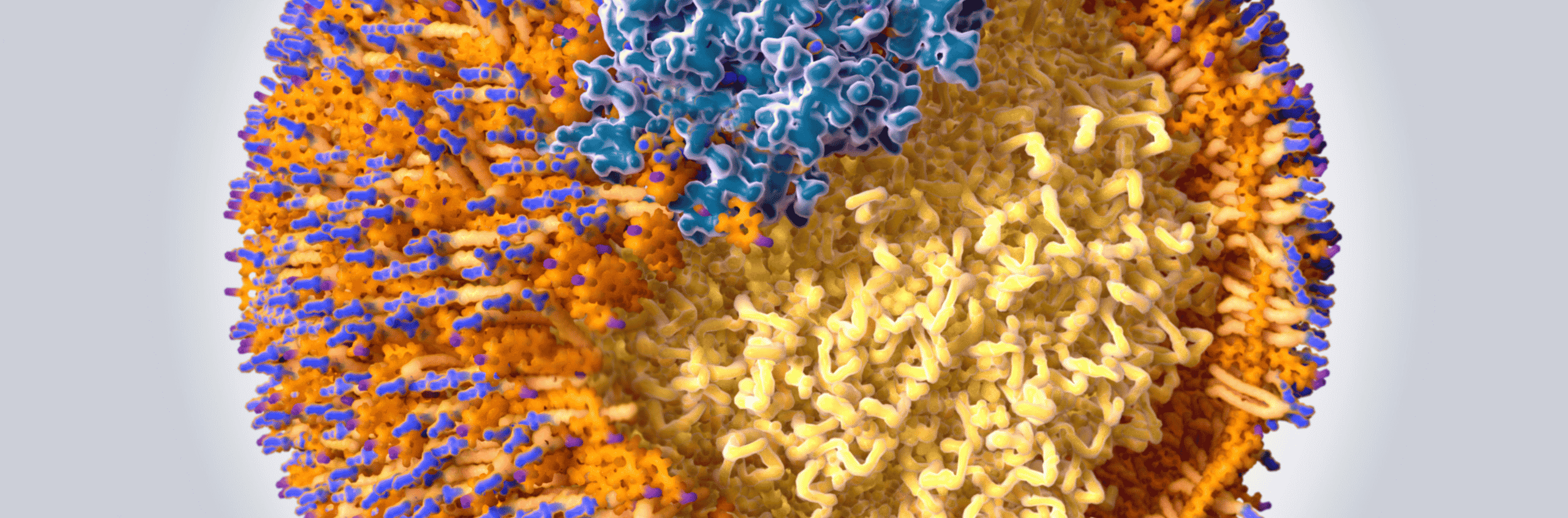
    Despite better modern management of risks like high blood pressure and cholesterol. Researchers suspected that there must be other things causing cardiovascular disease. Studies led to the discovery of a substance in the blood called lipoprotein (a). The level of lipoprotein (a) in the blood is different for everyone. Recent genetic studies proved that you inherit most of your high lipoprotein (a) levels. Your liver manufactures lipoproteins. They are a combination of fat and protein. Their job is to transport fats around the body in the blood. Lipoprotein (a) is a special combination, it includes a lipoprotein like LDL cholesterol, plus two more proteins, apolipoproteins A and B. The combination is very sticky and easily passes into the wall of the artery. This inevitably causes blockages of your blood vessels. So high lipoprotein (a) levels equal a high risk of cardiovascular disease.

    Even people with amazing lifestyles and diets can have high levels. If you are reading this then there is at least a 10 to 20% chance you have a high lipoprotein (a) level. The only way to know your level is through a blood test. Most people only need to check once in their lifetime. High levels mean you are at high risk for heart attacks and stroke. This is true even if your blood pressure and cholesterol levels are “ok”.

    Knowing you are at high risk we can then help you reduce that risk. We will look at all your cardiovascular risk factors and make a plan to optimise these. Knowing about a high lipoprotein (a) level can completely change treatment plans. We need to track your cholesterol measurements. The goals for your cholesterol need to be much lower to reduce your risk. This might mean using medicines to reduce targets like non-HDL cholesterol.

    Ongoing drug trials show great promise. In the future, drugs might directly reduce lipoprotein (a) levels.

    The European Society of Cardiology recommends measuring every adult’s lipoprotein (a) level. Unfortunately in 2022 the British Journal of Cardiology said “most clinicians are unaware of it”. We are passionate about the power of health screening to prevent disease. We recommend testing lipoprotein (a) levels for all adults.  Because levels are mostly genetically determined, it only needs to be checked once. This helps our patients prevent cardiovascular disease. Because prevention is better than cure.

    If you’re interested in learning more about lipoprotein (a) then check out Dr Hugh’s deep dive on lipids here. 

    British Society for Genetic Medicine  ·  European Atherosclerosis Society  · 
    Independent Doctors Federation

    As seen in BBC, The Guardian, Women’s Health, The Times and Tatler

    For educational purposes. This article is written by a practising GP and is intended to inform, not replace a consultation with your own doctor. It does not constitute medical advice. A note on clinical guidelines: Coyne Medical is an independent private clinic. Our approach is guided by the best available evidence, tailored to each individual. This may go beyond standard NHS or NICE guidance, which is designed for population-level care and weighted by resource constraints.
  • What is Apolipoprotein B and why is it important?

    What is Apolipoprotein B and why is it important?

    Atherosclerosis is the disease process that causes coronary artery disease. It is not a new condition. It has been found in 4000-year-old Egyptian mummies. In modern medicine, cholesterol is often described as being either good or bad. However, cholesterol is only ‘bad’ when it is in the wall of the artery. But how does it get there? And what makes it stay in the wall of an artery and cause a problem? Many things carry cholesterol around the body including HDL (often referred to as the good cholesterol) and red blood cells. Indeed, red blood cells carry a similar amount of cholesterol around the body as LDL particles (often referred to as the bad cholesterol).

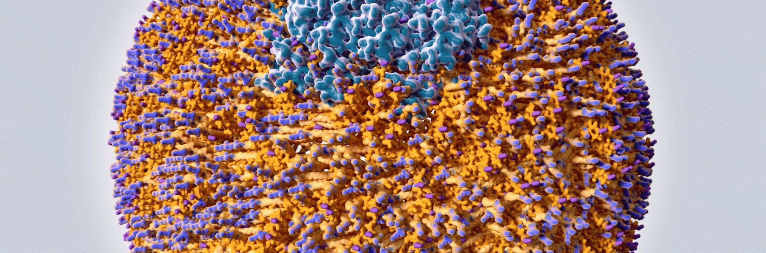
    So what is it that makes LDL bad? Or to put it another way, how does good cholesterol turn bad? The answer is Apolipoprotein B. If you look at the picture of an LDL particle such as the one above, you can see what it is made of. Fat is not soluble in water, so in the bloodstream, it must be transported in a packaged group. Each LDL particle consists of one Apolipoprotein B (ApoB), which organises other proteins, lots of cholesterol molecules and a core of polyunsaturated fatty acids. Think of each LDL as being like a group of tourists in a strange city being guided by a tour guide, the ApoB. 

    It is not only LDL that ApoB is present on. There is also one ApoB molecule on each intermediate-density lipoprotein (IDL), very low-density lipoprotein (VLDL) and chylomicrons. Indeed all of these particles also have the ability to cause cardiovascular disease (CVD). This is why a measure of all of these atherogenic particles, referred to as non-HDL cholesterol, is thought to be a better marker of cardiovascular disease risk than alone.

    The role of ApoB in atherogenesis, that is, the formation of atherosclerosis, is described by the “response to retention” hypothesis. Think of the artery as a road. The parts of the arteries that tend to have the most atherosclerosis are busy junctions. In cities the world over, busy junctions are often the sites of groups of bars and restaurants. Imagine now our group of tourists, the group of cholesterols with their ApoB guide wandering around the city. Imagine further that our group of tourists isn’t just any group of tourists but that they are a stag-do. Now a stag-do, our group of cholesterols, wants to find the busiest bar to go into. So it’s ApoB tour guide takes them to a bar at one of the busiest junctions and gets them in past the bouncer at the door. 

    Once inside the bar, the owner wants to keep this group in so that they continue to spend their cash in there. The artery lining acts in a similar way. But ultimately, acting in this way is going to be unhelpful for both the bar and the artery lining. How does the bar owner keep the group in the bar? Of course, by putting on the football, giving the tour guide a free drink and starting a happy hour. The artery lining acts, in the same way, to keep the LDL in the bar. Lipoprotein lipase and secretory sphingomyelinase are enzymes in the arterial wall that aid the retention of the ApoB and its cholesterol particles there. In the bar, as it’s getting busier and the punters are getting drunker our stag-do is swallowed up in the melee and drinks are being spilt everywhere. In the arterial lining, the ApoB and its cholesterols get swallowed up by macrophages (white cells) creating foam cells. Everyone in the bar is getting very merry, the atmosphere is really getting going, drinks are flying, people are up on the bar and more punters want to join the action. In the arterial lining, enzyme activation causes more retention of atherogenic particles. And it’s just then that the trouble really starts. As drinks and elbows start to fly and things get heated and people start to push past each other. First, a bottle smashes the window and then someone falls through the fire escape and everyone spills onto the street. The police are called. The area is blocked off. In the artery, the macrophages make enzymes that weaken the overlying cap and also release tissue factor. This agent is prothrombotic, it causes clots to form. It is the equivalent of calling the cops. Patry’s over. The artery is blocked.        

    So which is better at determining the risk of cardiovascular disease, non-HDL (which reflects the mass of cholesterol) or ApoB (which reflects the number of atherogenic particles)? To determine this requires studies of discordance. These studies show that when non-HDL was high but ApoB was normal, CVD risk was not high. But when non-HDL was normal and ApoB was high, CVD risk was high. Thus ApoB is a superior measure of CVD risk.

    So, we know that ApoB causes heart disease. New studies using a technique called Mendelian Randomisation have demonstrated that ApoB is implicated not only in heart disease but in stroke and diabetes. This type of study has also demonstrated that ApoB is associated with a significant reduction in lifespan. MR is a type of statistical analysis that uses variations in our genes to determine whether a certain exposure leads to a certain outcome. The fantastic thing about these studies is that confounding variables are removed. So, Mandellion Randomisation is excellent at establishing causality. In contrast, in an observational study causality can be difficult to determine because of confounding factors. A confounding factor is a variable that a study does not account for. An example of this would be a study looking at the link between heart disease and obesity that failed to take into account other important confounders such as age and smoking. Mendelian randomisation also eliminates reverse causality. This is a feature of observational studies when it can be difficult to ascertain what is the cause and what is the effect. An example of this would be does smoking cause lung cancer or does lung cancer cause smoking? 

    So when should we be measuring ApoB? The American Heart Association 2018 guideline on cholesterol management suggests that for people at intermediate risk of heart disease (risk between 7.5% and 20% on conventional risk scores) ApoB measurement can refine the selection of people at the highest risk. The 2019 European Society of Cardiology and the European Atherosclerosis Guidelines suggest that ApoB is the most accurate measure of CVD risk and a more accurate measure of lipid-lowering treatments. I mentioned previously the potential discordance between non-HDL and Apo-B. This is most common in people who are overweight, diabetic or have high blood values of triglycerides. These people should certainly have ApoB measured. However, atherosclerosis plaques grow over time. With the passage of time, more ApoB-containing particles are retained in the artery wall. So it is not just the number of ApoB-containing particles but the duration of exposure to them that is important. Therefore, it’s important to maintain low ApoB levels throughout life and start screening with ApoB early.  

    British Association of Sports and Exercise Medicine  ·  European Atherosclerosis Society  · 
    Independent Doctors Federation

    As seen in The Independent, The Daily Mail and Tatler

    For educational purposes. This article is written by a practising GP and is intended to inform, not replace a consultation with your own doctor. It does not constitute medical advice. A note on clinical guidelines: Coyne Medical is an independent private clinic. Our approach is guided by the best available evidence, tailored to each individual. This may go beyond standard NHS or NICE guidance, which is designed for population-level care and weighted by resource constraints.

  • Lipoprotein(a) – the bad guy of lipids

    Lipoprotein(a) – the bad guy of lipids

    In a previous blog, I tried to explain how dyslipidaemia (abnormal levels of blood lipids) causes atherosclerosis (plaques forming in the arteries) using the analogy of a stag-do. In this blog, I’m going to stretch this analogy even further. 

    In the stag do analogy, low-density lipoprotein (LDL) particles are members of the stag do. This is because it is LDL that causes all the trouble in the wall of the artery. In the same way, stag-dos cause trouble in bars the world over. The way to think of lipoprotein(a) is as the really bad, malevolent guy on a stag do. The guy with an evil streak that everyone else is a little bit wary of. 

    Lipoprotein (a) is a type of LDL particle. It is made up of a glycoprotein (a sugar linked to a protein), an apolipoprotein B molecule that is linked to an apolipoprotein (a). 

    Lipoprotein (a) isn’t all bad. It does have an important role in acute inflammation and wound healing. In our stag-do analogy, our protagonist can be thought of as a guy who is edgy, who brings a sense of excitement, a certain frisson. But this guy is one of those people who are best in small doses. On a stag-do, this bad dude is the type of person who would buy the stag 8 shots of gin to render him comatose, cheat on his partner, start a fight in a bar and get everyone kicked out. In the body, lipoprotein (a) causes problems by:

    • Interfering with the breakdown of clots.
    • Interfering with the functioning of the lining of the artery. 
    • Increasing inflammation in the wall of the artery.
    • Accelerating the formation of foam cells which leads to atherosclerotic plaque formation. 

    The main determinant of our levels of lipoprotein (a) is our genes. Levels can be measured using a standard blood test. Unfortunately, Lp(a) is not routinely measured on standard blood tests. Generally, the levels of Lp(a) remain stable over time although they can be affected to some extent by reduced levels of oestrogen and due to inflammation. 

    High levels of Lp(a) are associated with increased risk of heart and vascular disease as well as narrowing of the aorta. People who have a condition known as Familial Hypercholesterolaemia have a higher likelihood of having high levels of Lp(a). In these people, high Lp(a) further increases their already high risk of developing cardiovascular disease. 

    The European Society of Cardiology recommends that everyone should have their levels of Lp(a) checked at least once in their lifetime. Certainly, the following groups of people should have their Lp(a) levels checked:

    • Those with premature cardiovascular disease. 
    • People with a family history of premature cardiovascular disease (<55 years of age in men and <65 years of age in women).
    • People with a family history of elevated Lp(a).
    • People with a history of Familial Hypercholesterolaemia. 
    • People with a history of recurrent cardiovascular disease despite optimum treatment. 

    In addition, Lp(a) measurement can be useful for people with borderline cardiovascular risk scores who doctors are considering treatment with a statin and in people whose LDL cholesterol has not lowered as expected despite statin treatment. 

    So what can be done with elevated levels of Lp(a)? Currently, there is only limited scientific evidence to suggest that lowering Lp(a) reduced the risk of heart and vascular disease. Therefore, our focus of treatment is to reduce other risk factors for heart disease as much as possible. This includes lowering the levels of LDL as much as possible, usually with one or more medications. These medications include statins and another cholesterol-lowering medication called ezetimibe. 

    For some people, it is not possible to achieve optimum levels of LDL even with a statin at the maximum tolerated dose and ezetimibe, another cholesterol-lowering drug. For these patients, a medication called a PCSK9 inhibitor is required. These medications have been shown to reduce Lp(a) in some people and reduce the risk of heart disease independent of levels of LDL.  

    Some doctors use Nicotinic acid to reduce Lp(a). This medication has side effects that make it difficult to tolerate. Medical studies have shown that it does reduce Lp(a). However, these studies have not demonstrated that Nicotinic acid reduces the risk of heart disease. Another treatment called apheresis can reduce levels of Lp(a) temporarily but it is not clear yet whether this reduces the risk of heart disease. 

    A new promising therapy for elevated Lp(a) is antisense therapy. This is a type of gene therapy that intercepts the message from the body’s genetic code to make one of the key components of Lp(a). Trials in this treatment are ongoing. 

    Although there is no really effective current treatment to lower Lp(a) it is still very worth having levels checked at some stage of your life. Knowing your Lp(a) means that if they are elevated you can take steps that significantly reduce your risk of heart disease. 

    British Association of Sports and Exercise Medicine  ·  European Atherosclerosis Society  · 
    Independent Doctors Federation

    As seen in The Independent, The Daily Mail and Tatler

    For educational purposes. This article is written by a practising GP and is intended to inform, not replace a consultation with your own doctor. It does not constitute medical advice. A note on clinical guidelines: Coyne Medical is an independent private clinic. Our approach is guided by the best available evidence, tailored to each individual. This may go beyond standard NHS or NICE guidance, which is designed for population-level care and weighted by resource constraints.

  • Going Beyond ‘Cholesterol’

    Going Beyond ‘Cholesterol’

    When most people have their cholesterol level checked they will usually have their ‘total cholesterol,’ LDL-C (the concentration of LDL), HDL-C (the concentration of HDL), triglycerides (TG), and non-HDL measured. However, while these measurements are of some use in estimating the risk of cardiovascular disease they do not give the full picture. More important than the mass concentration of the cholesterol-containing particles is the number of cholesterol particles and more besides. 

    When most people think about cholesterol they think that there is a good type and a bad type. In fact, all cholesterol is good. Cholesterol is vital to our existence. It is one of the most important constituents of our cell membranes. Not only do cell membranes provide structural support for the cell but they also facilitate how cells move, what goes in and out of cells and how cells interact with each other. Cholesterol is also essential in the production of steroid hormones, vitamins and bile acids which help digestion. 

    So cholesterol is good. It is only bad when it is in the wrong place and the wrong place for cholesterol to be is in the arterial wall. But how does cholesterol get there. 

    Only about a quarter of the cholesterol in our bodies comes from what we eat. The rest is produced by our bodies. Our liver manufactures about a fifth of the cholesterol made by our bodies. Of the cholesterol that we eat, most of it is not absorbed by our gut. So the amount of cholesterol that we eat only has a limited effect on the cholesterol levels in the body. 

    I think of the process of coronary artery disease as being a bit like my stag do in Bratislava. This may seem like a stretch, but bear with me. Think of the guys on the stag do as being like cholesterol particles. Just like the members of a drunken stag do in an unfamiliar town, cholesterol needs something to take it around the body. The things that help transport cholesterol around the body are called apoproteins. These are like the group of girls from a tour operator that the best man hires to take the stag do on a tour of the town’s bars. In the bloodstream, the cholesterol particles and apoproteins when packaged together are called apolipoproteins. These cholesterol particles with their apoprotein chaperones (aka apolipoproteins) all bundle together. The group of cholesterol particles and its apoprotein chaperone is called a lipoprotein.

    Just like the group of girls from a stag-do tour operator, apoproteins help keep the group together and facilitate communication between the group and those outside the group. Apoproteins also help act as cofactors in enzyme reactions. This is akin to the female tour guides getting the stag group a round of free drinks when they arrive in a bar.  

    There are two important types of apolipoprotein – A and B. Apolipoprotein B (apoB) is almost always found with low-density lipoprotein (LDL). Think of low-density lipoprotein as a stag do with a female tour guide. Apolipoprotein A (apoA) is almost always found with high-density lipoprotein (HDL). Think of HDL as a tour group from a cruise ship with a nice tour guide. The are not going to cause trouble (unless the town is totally overrun with cruise ships and tour groups). An important point here, there is one ApoB molecule with one LDL particle. Each LDL particle is actually a group of cholesterol particles.  

    Most stag dos attract a few hangers-on. In the body, these unhelpful hangers-on are triglycerides. As the lipoproteins transport the cholesterol around the body these hangers-on drop away. Much the same happens on a stag do as it progresses.  

    The lining of an artery is called the epithelium. To cause atherosclerosis, i.e. cardiovascular disease, the LDL particle has to enter the endothelium. If you can imagine the lining of the artery as being like a strip of nightclubs and bars and the lumen of the artery as being like a road. The stag do, of course, are the LDL particles. Apoprotein B can get the stag do into the cool club. Each stag do needs to have a girl with them to get in (because there is there is one ApoB molecule with one LDL particle). In the same way, to get into the lining of the arterial wall, each LDL particle needs an Apolipoprotein B to chaperone past the bouncer on the door. In case you’re wondering, the bouncer in this instance is called NPC1L1Niemann-Pick C1-Like 1 or NPC1L1 for short. 

    So what is it that causes LDL to go into the lining of the artery? Remember, conventional blood tests for cholesterol measure the concentration of the different lipoprotein subfractions. But, it is the number of particles of LDL that is more important. Think of it this way. The more small stag do groups there are the easier it is for each stag do to get into a bar. No bouncer is going to let a massive number of drunk guys in one big group into a bar. Bigger stag dos with more people in each of them are less likely to get into a bar. In the same way the higher the number of LDL particles the greater the chance they have of getting into the wall of the artery. Although LDL concentration may correspond to particle number, often it doesn’t. 

    Indeed, it has been found that the best predictor of the risk of cardiovascular disease is LDL particle number. Furthermore, those people at the highest risk had a low (normal) LDL-C and a high LDL particle number. In medical statistics, when two variables that usually track together do not track together, it is known as discordance. People who have high LDL-C and high LDL particle number are also at high risk but not as high as those with discordant values. Thinking about it in stag-do terms, the highest risk of trouble comes when lots of small stag dos are let into a bar. In the opposite scenario if there is just one stag do allowed in but it is large with lots of people who know each other the chance of trouble is low. That is, if there is a high LDL-C and low LDL particle number the risk of cardiovascular disease is actually less. As I have mentioned, having a large number of LDL particles and a high LDL-C is also a high risk for cardiovascular disease. It’s just not as high risk as having low LDL-C and high particle numbers. Even in patients who show up to the hospital having a heart attack, frequently their LDL-C is normal.

    People with metabolic syndrome are more likely to have an unhelpful discordant relationship between LDL-C and LDL particle number. Metabolic syndrome is a condition in which people have raised triglycerides (a storage fat in the blood), raised blood pressure, elevated waist circumference and elevated blood sugar. 

    Once inside the club (the arterial lining) the stag do (LDL) causes all sorts of problems. Imagine members of the stag do hitting on local girls, being generally obnoxious and riling up groups of local guys. The same happens when LDL enters the endothelium – absolute pandemonium. This leads to inflammation in the arterial wall. On a stag do, the groups of locals might try to get rid of the stag do. But there are too many guys on the stag do. As you can imagine, the bar gets damaged. A brawl ensues and it spills out onto the street. Pretty much the same happens to the endothelium of the artery. The inflammatory response causes an arterial plaque to form which spills over into the lumen eventually causing a clot to form, blocking the artery. Think of the clot as the police being called. They then block off the street. This obviously means that bars and clubs further down the street have to close. This is equivalent to tissue ischaemia in our bodies where a lack of blood supply causes the tissues downstream to die. This is the process of a heart attack.  

    So although LDL-C can give us some information on the likelihood of having a heart attack, it is nowhere near as good as knowing the LDL particle number. Measuring this requires nuclear magnetic resonance spectrometry. This is very expensive. As there is one LDL per ApoB it turns out that ApoB is a good proxy for estimating LDL particle number and therefore cardiovascular risk. Indeed, several studies, including AMORIS, INTERHEART, IDEAL and the Leiden Heart Study have demonstrated that ApoB is superior in predicting cardiovascular disease risk. ApoB level is also useful in finding out how effective lifestyle changes and treatments have been. According to the INTERHEART and AMORIS trials, the best risk predictor of cardiovascular disease is the ratio between ApoB and ApoA1.  

    Ischaemic heart disease is, to a great extent, inflammatory. The real problem is not so much the presence of the LDL particle in the artery wall. It is the inflammatory response to the LDL that is the problem. LP-PLA is a marker of arterial inflammation and suggests that the formation of unstable plaques in the arteries is occurring. In our stag-do analogy, Lp-PLA is the equivalent of a group of bouncers entering the melee. It suggests that the arteries are angry and that arterial plaque is forming. The PLAC test measures the amount of lipoprotein-associated phospholipase (Lp-PLA2) in the blood. Lp-PLA is a key player in making plaques that line the arterial wall unstable and more likely to rupture.

    Another way of estimating the amount of inflammation in our arteries is with hs-CRP. Other issues can cause a rise in hs-CRP. However, in a patient who is at high risk of cardiovascular disease, I associate high levels of hs-CRP with being at very high risk for cardiovascular disease.

    So while traditional tests for cholesterol go some way to estimating cardiovascular risk, getting a much better estimation of risk requires a deep dive into different marke

    British Association of Sports and Exercise Medicine  ·  European Atherosclerosis Society  · 
    Independent Doctors Federation

    As seen in The Independent, The Daily Mail and Tatler

    For educational purposes. This article is written by a practising GP and is intended to inform, not replace a consultation with your own doctor. It does not constitute medical advice. A note on clinical guidelines: Coyne Medical is an independent private clinic. Our approach is guided by the best available evidence, tailored to each individual. This may go beyond standard NHS or NICE guidance, which is designed for population-level care and weighted by resource constraints.